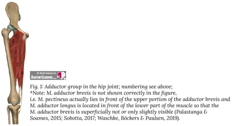
The term adductor muscles (comes from the Latin “adducere” ≙ “to bring up”) in general sport-related or medical usage usually refers to a group of muscles that are located on the inside of the thighs. Of course, there are also adductors or muscles that act as adductors in other parts of the body. This article is all about the adductor group, which acts on the hip joint. Probably the most important adductors that belong to this group of adductors are the main topic of this article and are listed below and shown in Figure 1.
Adductor muscles – Anatomy
(1) M. adductor magnus (lat. „magnus“ ≙ „big“)
(2) M. adductor longus (lat. „longus“ ≙ „long“)
(3) M. adductor brevis (lat. „brevis“ ≙ „short“)
(4) M. gracilis (lat. „gracilis“ ≙ „slim“ oder „thin“)
(5) M. pectineus (lat. „pectineus“ derives from „pecten“ ≙ „ridge“)
The following muscles may also help with adduction:
M. obturatorius externus; M. quadratus femoris; M. gluteus maximus
Adductor muscles – M. adductor magnus
If you look at the front of the thigh, you can only partially see the adductor magnus (see Fig. 1). The muscle is extended towards the surface by the Mm. adductor brevis and longus obscured. The adductor magnus muscle lies in front of the hamstrings (specifically in front of the semimembranosus and semitendinosus muscles). The muscle has an extensive origin, ranging from the pubic ramus (“ramus inferior ossis pubis”) to the lower area on the outside of a rough bony prominence on the ischium (“os ischii” ≙ “ischial bone”; “tuber ischiadicum” ≙ the named “ bony prominence”).
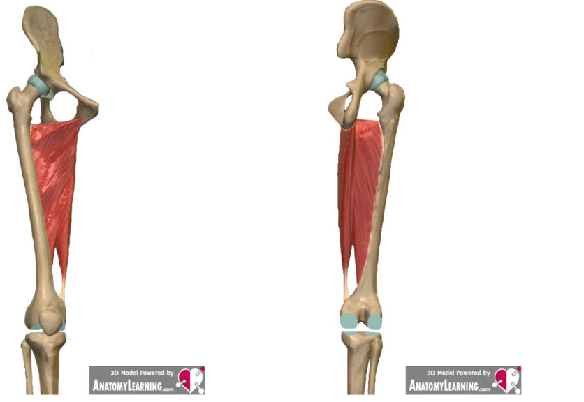
As can be seen in Fig. 2 and 3, the muscle has different courses. The part of the muscle that originates in front of the pubic ramus runs outwards and a little backwards on the way to its insertion, in order to then attach on the one hand on the middle area of a bony ridge on the thigh bone and on the other hand also almost horizontally on the thigh (“Linea aspera” ≙ the bony crest mentioned; “Labium mediale” ≙ the middle area or the “middle lip” of the Linea aspera) (Jeno & Schindler, 2018; Palastanga & Soames, 2015).
While the portion of the adductor magnus that arises from the pubic rami anteriorly rotates to then insert on the femur, the posterior portion of the muscle that arises from the ischium pulls straight down to insert at a bony prominence on the internal condyle of the femur (see Fig. 2 and 3; “Epicondylus medialis femoris” ≙ the mentioned condyle; “Tuberculum adductorium” ≙ the mentioned bone elevation). It is possible that some fibers of the muscle radiate into the medial ligament (“ligamentum collaterale mediale”) of the knee joint (Palastanga & Soames, 2015). A small and incomplete, not present in all adults (Tubbs et al., 2011), near the pelvis separation of the adductor magnus is called “M. adductor minimus” (Sobotta, 2017; Waschke, Böckers & Paulsen, 2019). When looking at Figures 2 and 3, it is noticeable that the area between the two attachments of the adductor magnus forms a kind of opening. This is the so-called “hiatus adductorius”. Pass through this opening u. the large femoral artery and the large femoral vein (Waschke, Böckers & Paulsen, 2019; called artery ≙ femoral artery; called vein ≙ femoral vein).
Adductor muscles – M. adductor longus
As described above, the adductor longus lies in front of the adductor magnus and partially covers it. It originates at the front of the pubic bone (“os pubis”) and travels down and outwards to the inner thigh to also attach to the bony crest “linea aspera” behind the inner head of the quadriceps (the inner or medial head of the quadriceps is the “vastus medialis “).
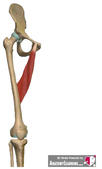
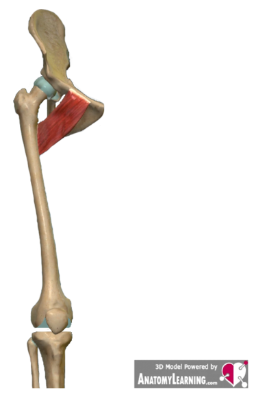
Adductor muscles – M. adductor brevis
The origin of this short muscle is at the front, somewhat to the side, on the pubic bone and on the lower branch of the pubic bone (see Fig. 5; “pubic bone” ≙ “os pubis”; “lower branch of the pubic bone” ≙ “ramus inferior ossis pubis”). The adductor brevis runs diagonally downwards from the pubic bone to attach to the upper half of the above-mentioned to attach the bony ridge of the thigh. Looking at the thigh from the front, the muscle is covered by the pectineus muscle above and the adductor longus muscle below.
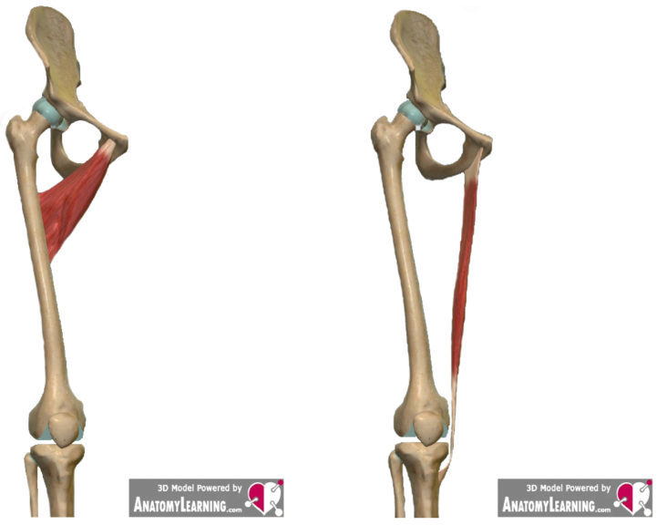
Adductor muscles – M. gracilis
The gracilis (Fig. 6) is a long, thin muscle that extends from the front of the pubic bone and down from the inferior pubic ramus to attach on the inside of the tibia below the internal condyle (“pubic bone” ≙ “os pubis”; “inferior pubic ramus” ≙ “ramus inferior ossis pubis”; “inner articular condyle of the tibia” ≙ “condylus medialis tibiae”).
Essentially, the Gracilis shares a common approach with the Mm. sartorius and semitendinosus. This collective attachment area is also referred to as “pes anserinus superficialis” (Sobotta, 2017; Waschke, Böckers & Paulsen, 2019; due to the 3 tendons involved, the joint attachment is reminiscent of a goose foot → “pes” ≙ “foot”; “anserinus” ≙ “Goose”; “Superficialis” ≙ “superficial”).
Adductor muscles – M. pectineus
The pectineus originates relatively widely. Its origin is on a bony edge on the upper pubic ramus, on a bony tubercle of the pubic bone and at a connection point between the pubic bone and ilium (the above-mentioned “bone edge” ≙ “Pecten ossis pubis”; “upper pubic ramus” ≙ “Ramus superior ossis pubis”; the above-mentioned “bony tubercle” ≙ “Tuberculum pubicum”; the “connection point between pubic bone and ilium” ≙ “Eminentia iliopubica”). At its base it pulls backwards down and outwards to the thigh. On the upper thigh it radiates into a bony ridge (the so-called “bony ridge” ≙ “Linea pectinea femoris”) (Palastanga & Soames, 2015).
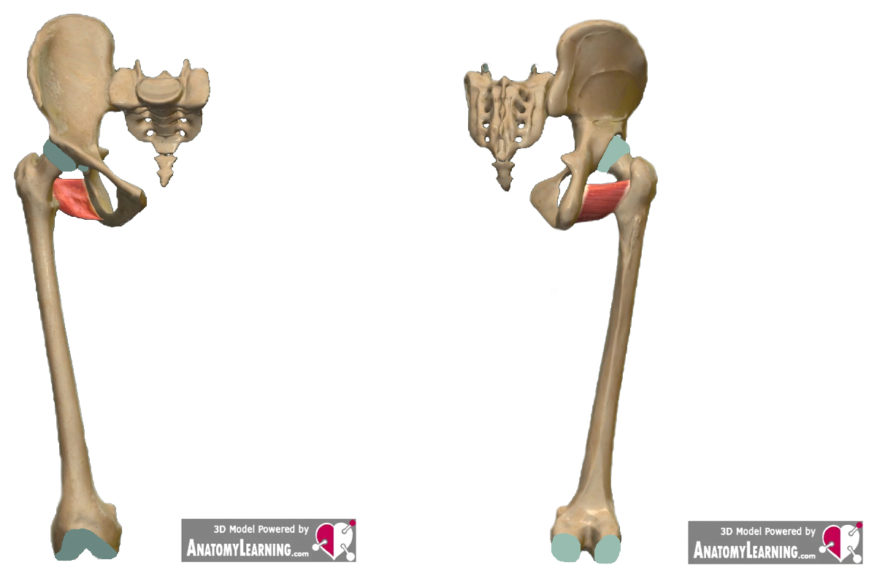
Adductor muscles – M. obturatorius externus
Behind the Latin name one can assume the verb “obturare”, which can be translated as “to clog”. A noun for this would be e.g. “seal”. “Externus” indicates “externally”. Accordingly, it can be assumed that the muscle seals something. And indeed, the muscle lies over an opening in the pelvis, the “foramen obturatum”.
The outer surface of a membrane between the pubic bone and the ischium as well as its bony edges serve as the origin of the obturatorius externus (the so-called membrane ≙ “membrana obturatoria”), and the muscle uses a depression on the back of the femoral neck as an attachment (the so-called depression ≙ “fossa trochanterica “).
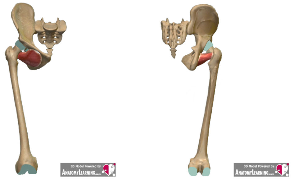
Adductor muscles – M. quadratus femoris
The Latin name already indicates the shape and position of the muscle (Figs. 10 and 11). This square muscle arises from a rough bony prominence on the ischial bone (called the bony prominence ≙ “tuber ischialicum”). It pulls outwards towards the thigh (“femur”) in order to start in an elevation of the groin between the two trochanters (the groin ≙ “crista intertrochanterica”; the elevation ≙ “tuberculum quadratum”; the lesser trochanter ≙ “trochanter minor”; the large trochanters ≙ “trochanter major”).
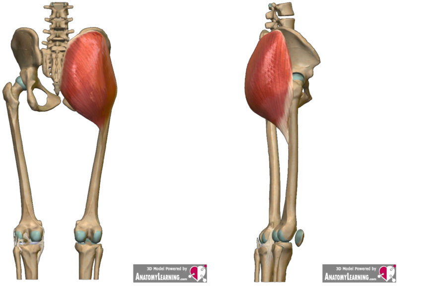
Adductor muscles – M. gluteus maximus
As can be seen in Figures 12 and 13, the gluteus maximus has a broad origin. Essentially, it originates from the iliac bone, sacrum, from a ligament in the rear part of the pelvis and a large fascia in the lumbar region (the origins are a little more precise: “Facies glutea” on the “Os ilium”; “Facies posterior”; “Lig. Sacrotuberale”; thoracolumbar fascia). The cranial portion of the muscle pulls outward and down to radiate onto a tough strand of connective tissue on the outside of the thigh as part of a large fascia that surrounds the muscles of the thigh. The caudal part of the gluteus maximus attaches to a rough area between the greater trochanter and a ridge of bone on the thigh (the connective tissue cord called ≙ “iliotibial band”; the called fascia ≙ “fascia lata”; the rough area ≙ “tuberositas glutea”; the Bar ≙ “Linea aspera”) (Waschke, Böckers & Paulsen, 2019).
Adductor muscles – Functions
The muscles of the adductor group primarily live up to their name and adduct in the hip joint. That is, the thigh is brought to the center of the body (in the frontal plane). According to Palastanga & Soames (2015), the adductors are strongest in the “anatomically normal position (nullposition)”*.
Examples:
You have a ball between your legs and you are trying to crush the ball. The movement may not be that big or not obvious at first glance, but the adductors are the main driver behind this action.
You lie on your back and your legs are stretched perpendicular to the ceiling. This is position 1. Now you spread your legs wide apart (like a splits, just… well… different 😊) and hold the position. That’s position 2. And now, when you bring your legs back from position 2 back to position 1, it’s essentially the adductors that make it possible.
Functional activity
In addition to adduction as the main function of the adductor group, the muscles also help to carry out other movements. The adductors stabilize the pelvis when standing on one leg (Waschke, Böckers & Paulsen, 2019) and when walking (Sobotta, 2017; Waschke, Böckers & Paulsen, 2019). Waschke, Böckers & Paulsen (2019) state that essentially all muscles of the adductor group are involved in flexion and external rotation in the hip joint because they cross the front of the hip joint, lie in front of the transverse axis and pull towards the posterior of the thigh (which causes external rotation According to Hochschild (2016) and Palastanga & Soames (2015), the M. gracilis does not seem to play a role; the part of the adductor magnus that originates on the ischium according to Palastanga & Soames (2015) and Waschke, Böckers & Paulsen (2019) not involved in hip flexion but aids in hip extension).
It may also not be entirely clear whether the adductor longus and adductor magnus muscles are more involved in internal or external rotation. It is possible that the functioning of at least the adductor magnus depends on the thigh position (Palastanga & Soames, 2015).
The M. gracilis has two joints and accordingly also acts on the knee joint by supporting knee flexion and internal rotation (Palastanga & Soames, 2015; Waschke, Böckers & Paulsen, 2019). The external obturator, quadratus femoris, and gluteus maximus muscles may also assist in adduction. In the gluteus maximus, adduction is performed through the caudal portion (Waschke, Böckers & Paulsen, 2019). The primary functions of the gluteus maximus are to extend and externally rotate the hip joint, particularly from a flexed position. In addition, this muscle can also aid in hip abduction and knee extension through its attachment to the iliotibial tract. The attachment to the iliotibial band also has a stabilizing effect on the outside of the knee. In addition, the gluteus maximus plays an important role in pelvic stabilization while standing and walking (Palastanga & Soames, 2015).
* Normal anatomical position → standing upright, face forward, arms hanging at sides, palms facing forward (or to body), legs side by side (hip-width apart), feet pointing straight ahead (Sobotta, 2017)
References
- Hochschild, J. (2016). Functional anatomy for physical therapists. Thieme.
- Jeno, S. H., & Schindler, G. S. (2018). Anatomy, bony pelvis and lower limb, thigh adductor magnus muscles. In StatPearls [Internet]. StatPearls Publishing.
- Palastanga, N., & Soames, R. (2015). Anatomie und menschliche Bewegung: Strukturen und Funktionen. Elsevier, Urban & Fischer Verlag.
- Sobotta, J. (2017). Sobotta, Atlas der Anatomie Band 1: Allgemeine Anatomie und Bewegungsapparat. Deutschland: Urban & Fischer in Elsevier.
- Tubbs, R. S., Griessenauer, C. J., Marshall, T., Dennison, C. P., Shoja, M. M., Loukas, M., … & Cohen-Gadol, A. A. (2011). The adductor minimus muscle revisited. Surgical and radiologic anatomy, 33(5), 429-432.
- Waschke, J., Böckers, T. M., & Paulsen, F. (Eds.). (2019). Sobotta Lehrbuch Anatomie. Elsevier Health Sciences.
Annotation:
If the source of the information is not specified separately in the text, then the findings in each of the above Sources 3, 4, 6 can be found.
The anatomical terms and the non-subject-specific prefixes are from Sobotta (2017).
The anatomical illustrations were created with Anatomy Learning: https://anatomylearning.com/
Be the first to comment